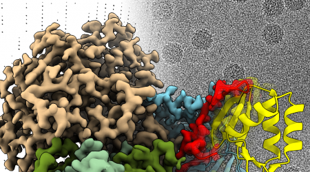
Bioimaging and microscopy
Bioimaging is a state-of-the-art microscopy facility supporting world-class science at the John Innes Centre. The platform offers a wide range of imaging applications in both light and electron microscopy. The team can offer a comprehensive service, including imaging and custom sample preparation.
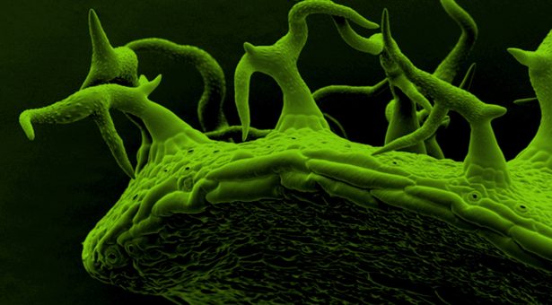
Training and technical support in a variety of microscopy techniques is available, including confocal microscopy, super-resolution techniques, scanning electron microscopy (SEM) and high-resolution transmission electron microscopy (TEM). The Bioimaging facility specialises in the imaging of plants and microbes offering assistance and advice on experimental design and image analysis. With more than 20 microscopes and many years of expertise, John Innes Centre’s Bioimaging team will give you an edge over the competition by providing the highest-quality images.
All external work has to be agreed on a case by case basis, depending on availability and capacity. Quotes will be provided upon request. For more information email Jake Richardson.
Techniques available
Light microscopy
- Brightfield, DIC (Normaski), phase (100x objective only), darkfield, polarised light and fluorescence imaging
- Laser scanning confocals with FRAP, FRET, FLIM and FCS capabilities
- To go beyond the diffraction limit, we also have a Zeiss LSM 980 Airyscan 2 and a Zeiss LSM 880 with Airyscan fast and a Zeiss Elyra 7 for SIM
Electron microscopy
- TEM (transmission electron microscope) and two SEM’s (scanning electron microscopes) and specialist sample preparation equipment
- Sample preparation by a variety of techniques for TEM, such as high-pressure freezing and freeze-substitution, resin embedding and sectioning, immuno-gold labelling, cryo-grid plunging or negative staining
- 200kV TEM for electron tomography, with high-tilt and dual-tilt holders, and cryo-TEM
- SEM – we offer critical point drying or cryo-SEM techniques. Both our SEMs have field-emission guns, giving the highest resolution capabilities. We also have the ability to do the volume EM techniques of serial block face SEM and array tomography.
Light microscopy equipment
Zeiss LSM 980 confocal with Airyscan 2
A point-scanning system on an upright Z.2 microscope with a motorised stage and a variety of objectives (air/oil/water/dipping). Available laser lines are: 405, 445, 488, 514, 561, 594, 639 nm. The system has two standard PMT detectors (tuneable), a more sensitive spectral detector array and an Airyscan 2 detector with multiplex mode for either super-resolution imaging or fast imaging.
Zeiss LSM 880 confocal with Airyscan fast
A point-scanning system on an upright Z.2 microscope with a motorised stage and a variety of objectives (air/oil/water/dipping), also suitable for DIC. Available laser lines are 405, 458, 488, 514, 561, 594, 633 nm. The system has two standard PMT detectors (tuneable), a more sensitive spectral detector array and an Airyscan detector for either super-resolution imaging or fast imaging.
Leica Stellaris 8 FALCON confocal
A point-scanning system on a fully motorised Leica DM6 upright microscope equipped with a variety of objectives (air/oil/water/dipping) also suitable for DIC. This microscope can be used for fluorescence lifetime imaging (FLIM) and fluorescence correlation spectroscopy (FCS). Available laser lines are 405, 488, 514 and 561 nm and a pulsed white light laser (440-790 nm) with pulse picker. Fluorescence detection is tunable (410-800 nm) with two HyD S and two HyD X detectors as well as two external APDs. The microscope has a standard and a resonance (fast) scanner. It is equipped with a heating/cooling stage insert.
Leica TCS SP8X confocal
This point-scanning system is based around a fully motorised Leica DM6 upright microscope equipped with a variety of objectives (air/oil/water/dipping) also suitable for DIC. Fluorescence detection is tuneable (400-800nm) with up to five internal (PMT plus HyDs x4) and two external detectors (SPADS). Available laser lines are 405, 440 (pulsed), 457, 488, 514, 561 nm and a pulsed white light laser tuneable from 470 – 670 nm. The scan head has two scanners: conventional (standard) and resonance (fast dynamic). The microscope can be used for fluorescence lifetime imaging (FLIM) and fluorescence correlation spectroscopy (FCS).
Zeiss Elyra 7
This microscope uses structured illumination microscopy (SIM) to break the diffraction barrier including SIM2 reconstruction for further resolution improvement. It can also be used in Apotome mode as a quasi-confocal when larger fields of view are required or as a sensitive widefield microscope with total internal reflection fluorescence (TIRF) capability. Built on an inverted Observer Z1 microscope with motorised stage, heated incubator, a variety of objectives and two PCO edge 4.2 sCMOS cameras for simultaneous two-colour imaging. Available laser lines are 405, 488, 561, 642 nm.
Zeiss Elyra PS1
This system uses super-resolution imaging methods that break the Abbe resolution limit: 3D structured illumination (SIM) and 2D single molecule localisation techniques (dSTORM and PALM). It is also capable of Total Internal Reflection Fluorescence (TIRF) and can be used as a highly sensitive wide-field system. Built on an inverted Observer Z1 microscope with motorised stage, incubator with temperature control and two EMCCD cameras, one of them back-thinned. Available lasers are 405, 488, 561 and 642 nm.
Zeiss Axio Observer Z1
A high-quality, inverted, wide-field microscope with motorised stage control (for z sectioning, time lapse, tiling), auto focus and a microscope incubator (range from RT to 37 degrees celsius) making it ideal for live cell imaging. Equipped with both colour and monochrome cameras. Imaging modalities are bright field, DIC (x100 only), phase contrast (x100 only) and fluorescence over a wide range of wavelengths. The fluorescence illumination is a Zeiss Colibri 7 LED light source giving independent control over 7 LED bands. Additionally, it has a RAPPA UGA42 laser targeting system for FRAP and photo-activation.
Zeiss Axio Imager Z2
This system is equipped with both colour (histology) and monochrome (fluorescence) cameras. The microscope stand is upright with motorised stage control (z sectioning, time lapse, tiling and multiple positions), software auto focus. Acquisition modalities are brightfield, DIC, phase contrast (100x objective only), darkfield (up to 40x objective), polarised light and fluorescence (complete visible range). The fluorescence illumination is a Zeiss Colibri 7 LED light source giving independent control over 7 LED bands.
Zeiss Axio Zoom V16 Fluorescence Stereo Microscope
Stereo microscope with automated stage, two Orca Flash4 sCMOS cameras for simultaneous time lapse imaging of two fluorescence channels and a Zeiss Axiocam 512 colour camera. Monocular z-sectioning and Apotome 2 for optical sectioning. Fluorescence illumination provided by Lumencor Spectra 3 LEDs with filter cubes for DAPI, CFP, GFP, YFP, RFP and dual bandpass filters. Brightfield via LED light ring or transmitted light with darkfield and oblique illumination options.
Leica M205FA Fluorescence Stereo Microscope
This microscope is computer controlled and can capture images using a Leica DFC310FX colour camera. Imaging modalities are reflective bright field (LED light ring), transmission illumination (bright field, dark field and Rottermann contrast) & fluorescence (LED illumination, for GFP and DSR). It can also perform time lapse.
Electron microscopy equipment
FEI Talos F200C TEM – A 200 KV transmission electron microscope with an FEG filament (field emission source) and a Gatan OneView camera; a 4k by 4k CMOS bottom-mounted camera. To support cryo-TEM, we have two Gatan Elsa holders and we are about to add a Falcon 4i direct electron detector. We will also have micro-ED capability via a Ceta-D camera. High and dual-tilt holders are available for electron tomography.
FEI Nova NanoSEM 450 – A high-resolution, field emission scanning electron microscope with a Gatan cryo-system, EDS, BSE and STEM detectors. Low voltage (1kV) resolution is 1.4 nm in high vacuum mode. It offers beam deceleration, both high and low vacuum modes, and is the perfect all-round tool for a wide range of samples.
Zeiss Gemini 300 SEM – A second, high-resolution, field emission scanning electron microscope with a Quorum cryo system. In addition to the standard secondary electron detector, there is an in-lens SE detector for high resolution work, a back scatter detector, and a Gatan OnPoint detector to complement the Gatan 3View system which is used for serial block face imaging. We can also perform array tomography using this SEM.
Vitrobot – for cryo-grid plunging in liquid ethane, for cryo-TEM.
Pelco EasiGlow – for glow discharging grids to make them more hydrophilic for TEM.
Leica ACE 200 coater – A carbon thread coater / glow discharge unit, with quartz crystal thickness monitoring and automated functions, for putting a homogenous, conductive layer of carbon on SEM samples for X-ray microanalysis or for glow discharging TEM grids.
SEM sample preparation
- Sputter coater – A Leica ACE600 high resolution sputter-coater for use with our field emission SEMs. Targets routinely available are Au or Pt.
- Critical Point dryer – A Leica CPD300 used for drying chemically fixed samples for the SEM.
Sectioning facilities
- Two Leica ultramicrotomes for ultra-thin and semi-thin resin sectioning for both light and electron microscopy applications. The UC7 is available for general use and, with a camera and monitor, is ideal for training purposes. The ARTOS 3D has automated serial sectioning capability for array tomography applications.
- Leica TRIM2 trrimming tool, to assist in cutting the perfect pyramid before sectioning
- Leica KMR3 knife-maker, for preparing glass knives, hot-plates for drying slides, and light microscopes for checking sections, along with a slide-staining area.
- A vibratome and a cryostat for light microscopy sectioning.
High pressure freezer and freeze substitution unit
A Leica HPM100 high-pressure freezer, with 3mm and 6mm carriers, used for freezing very small samples at pressures of ~2000 bar, to prevent ice-crystal growth. Sample size is extremely limited, but it can provide the best method for membrane-preservation. Due to the challenging nature of this technology, it requires technical assistance.
The Leica AFS2 for freeze substitution machine is generally used after high-pressure freezing but can also be used for the PLT embedding method (progressively lowering temperature). Both these methods have been shown to help retain antigenicity and are therefore used when immuno-gold labelling studies are required for TEM.
Tissue processor
The Leica EM TP embedding machine is used for automatically performing tissue dehydration and resin-infiltration prior to polymerisation of resin. We routinely use it for samples for electron microscopy, embedding in LR White resin. This machine is generally only used by Bioimaging staff but can be accessed if necessary.