Streptomyces bacteria key to understanding cell-division
Cell division is a fundamental process in all domains of life, required for the regeneration of damaged tissue, organ growth and development.
If cell division gets out of control, it can lead to cancer (you can find out a bit more about this in our blog – Controlling Cell Division: Do plants get cancer?).
In less complex organisms, like bacteria, cell division is important for reproduction and survival. The group of Dr Susan Schlimpert here at the John Innes Centre use the soil bacteria Streptomyces, famous for its earthy smell and the production of antibiotics, as a scientific model to understand cell division.
A recent study reveals new insights into this fundamental biological process in Streptomyces. First author Dr Matt Bush answers some questions about this research.
Why use Streptomyces to study cell division?
Much of our understanding of the mechanisms that underpin cell division in bacteria has been driven by studies using classical model organisms such as Escherichia coli and Bacillus subtilis. In recent years however, research from labs using other, non-classical organisms has revealed the tremendous diversity in the regulation and cell biology of division among microbes.
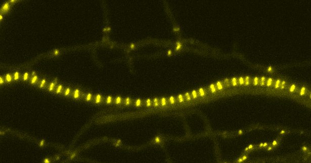
Streptomyces are a case-in-point. They are special when it comes to cell division because unlike most bacteria that simply split in half to produce two identical daughter cells, Streptomyces grow as long branched filaments (hyphae) containing many copies of the chromosome each of which needs to be separated and distributed into spores during reproduction.
Intriguingly, Streptomyces undergo two distinct forms of cell division:
- they synthesise so-called “cross-walls” or division septa that divide the growing filaments at irregular intervals to compartmentalise the hyphae. The individual filaments remain physically connected.
- they can undergo a process called sporulation in which the tip compartment is segmented into dozens of small (~1micron = 1e-4 cm) and equally sized spores that can eventually be dispersed into the environment to colonise new environments. The formation of spores is an important survival strategy for bacteria as they are able to tolerate prolonged harsh environmental conditions such as excessive heat or cold or a lack of nutrients.
What did your study find?
Why Streptomyces need “cross-walls” was previously unknown.
Our study identified a gene sepX, encoding a component that is essential to make cross-walls and this allowed us to functionally dissect their biological importance.
We found that cross-walls provide Streptomyces with an important fitness advantage: the compartments that form as a result of cross-walls protect the colony from complete collapse and cell death when there is a leak/puncture in the cell envelope. Furthermore, they are likely to be required for the compartment-specific accumulation of molecules that coordinate growth and development that eventually leads to sporulation and continued survival.
What are the next steps for this research?
The more we study new proteins involved in cell division, the deeper our understanding of this complex biological process. Our identification of SepX and the importance of cross-walls in Streptomyces raises additional questions that we may be able to address in the future.
Do cross-walls allow the exchange of signals and nutrients between neighbouring compartments in the colony? Such cell-to-cell communication channels are prevalent in plants and animals, suggesting that they may play an important role in bacteria like Streptomyces too.
See the full study in Nature Communications