Seeing the bigger picture: imaging technique widens our view on the inner worlds of plants and their guests
Plants come in all shapes and sizes – but why, and how?
Scientists at the John Innes Centre exploring how interactions between genes affect plant patterning have developed an imaging technique to visualise live gene activity at the macroscopic scale.
Why does a rose flower have layered petals, while daffodils have trumpets? Why is the shape of an oak leaf so different from that of a pine needle? Questions like these fascinate Professor Enrico Coen, whose research explores the development of patterns and shapes in plants.
To help answer these questions, Professor Coen’s team is developing novel imaging methods that allow researchers to look into the 3D depths of plant structures.
Recording plant structures started with botanical illustrations and then moved to photography. More recently, 3D methods such as optical projection tomography (OPT) allow us to see the gene activity inside living plants in 3D. The images can be used to build up a picture of how gene activity changes over time, which in turn can be fed into computer models to simulate and predict plant patterns.
However, until now, this technique was only good enough to visualise very small structures up to 1.5 cm across.
In a new paper published in Journal of Experimental Botany, Professor Coen and his team of cell and developmental biologists at the John Innes Centre describe a vast improvement on the existing OPT technique.
Karen Lee, first author of the paper and a research assistant in Professor Coen’s lab said: “We’ve successfully developed ‘Macro OPT’ (M-OPT) – a larger scale version of OPT that allows us to explore 3D gene activity in fixed or living plant structures up to 6 cm in length. As well as greatly increasing the diversity of plant structures we can study, this advance also helps us to better assess how gene activity in particular tissues affects the wider organism.”
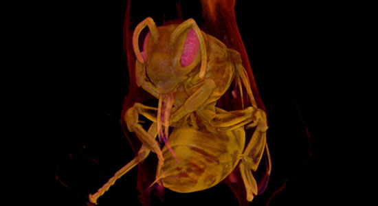
“A further exciting benefit is that we can also use M-OPT on animal tissues – so, for the first time, we have been able to visualise pollinating insects within flowers in 3D. In future, we could use M-OPT to see, for example, how pests damage leaves or roots, or precisely how a Venus fly trap changes shape to enclose and digest a trapped insect.”